X-ray imaging remains a fundamental first-line investigation in the assessment of foot and ankle pain. While not all patients require imaging, understanding when to refer for radiographs is essential in identifying structural pathology, guiding management, and avoiding missed diagnoses.
X-rays are particularly useful in evaluating bony structures, joint alignment, and degenerative change. Common indications include suspected fractures, assessment following acute injury, persistent pain where structural pathology is suspected, and evaluation of joint space narrowing or deformity. In the context of trauma, clinical decision rules may guide initial imaging, but ongoing symptoms or atypical presentations should prompt reconsideration.
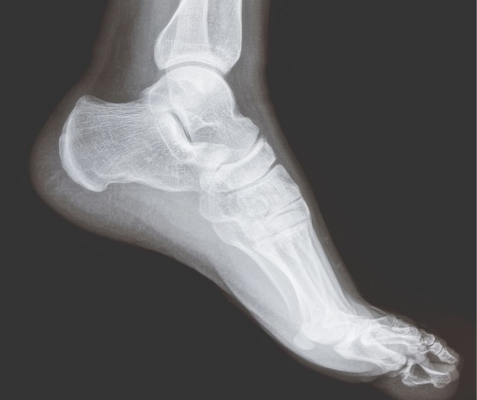
In non-traumatic cases, X-rays can provide valuable information in conditions such as osteoarthritis, hallux valgus, hallux rigidus, and midfoot degenerative change. Weight-bearing radiographs are especially important in the foot and ankle, as they allow assessment of functional alignment and load distribution, which may not be evident on non-weight-bearing images.
Initial management may not always require imaging, particularly in clearly defined soft tissue conditions responding well to conservative care. However, referral should be considered where symptoms persist despite appropriate management, where there is diagnostic uncertainty, or where structural pathology is suspected based on clinical assessment.
X-rays also play a role in excluding more serious pathology. Stress fractures, although sometimes not visible in early stages, may become apparent on follow-up imaging. Inflammatory or systemic conditions may demonstrate characteristic joint changes, and radiographs can provide a baseline for monitoring progression.
It is important to recognise the limitations of X-ray imaging. Soft tissue structures, including tendons, ligaments, and nerves, are not well visualised, and normal radiographs do not exclude significant pathology. Where clinical suspicion remains high despite normal X-rays, further imaging such as ultrasound or MRI may be required.
Multidisciplinary team (MDT) working enhances the value of imaging. Collaboration between clinicians, radiologists, podiatrists, orthotists, and orthopaedic surgeons ensures that imaging findings are interpreted within the clinical context and integrated into a coordinated management plan.
Referral for X-ray should therefore be considered as part of a broader clinical assessment, rather than in isolation. Used appropriately, it provides a valuable tool in identifying structural abnormalities, guiding treatment decisions, and supporting timely referral to specialist care when required.
For clinicians, understanding when to request X-ray imaging helps avoid both over-investigation and missed pathology. For patients, appropriate use of imaging supports more accurate diagnosis and a more structured approach to management.
: A Practical Guide for Clinicians")
